Radiation Safety Protocols
- September 20, 2024
- 3.5k views
- 4 min read
Radiography is an essential medical diagnostic tool that provides vital information about internal structures within the human body. However, ionizing radiation poses potential hazards in radiography, hence the call for strict safety procedures to protect patients and healthcare workers.
In the following section, we will explore some aspects of radiation safety protocols that should be in place in a radiography unit, encompassing principles, practices, and safety measures.

Table of Contents
Radiation Safety
ALARA stands for “As Low As Reasonably Achievable.” This is one of the cardinal principles of radiation protection in radiography. It protects the patient, the radiologic technologist, and other healthcare workers from ionizing radiation. Keep radiation exposure as low as possible—just enough to produce the desired effect while keeping it below any possible harmful level and realistically attainable.
Critical Elements of a Radiation Safety Protocol
Give an Explanation of Procedures
Justification for any radiographic procedure should precede it. The procedure’s benefits should outweigh the possible risks of radiation exposure. Limit unnecessary or redundant imaging and consider alternative techniques when ionizing radiation is not required.
Optimization of Exposure
Optimization is defined as the adjustment of radiation dose at a level as low as is reasonably achievable with conventional imaging methods; this can be performed in several ways:
- Technique Charts: These are pre-set technique charts that define the best exposure parameters, the type of exam, and patient size.
- Collimation: This reduces the X-ray scope to just the area of interest, thereby minimizing radiation dosage as much as possible to the surrounding tissues.
- Filtration: It filters out low-energy X-rays that can only increase patient dosage without contributing to image information.
- AEC: Automatic Exposure Control is the system of adjustment of exposure parameters based on patient anatomy and tissue density.
Protection Measures for Patients
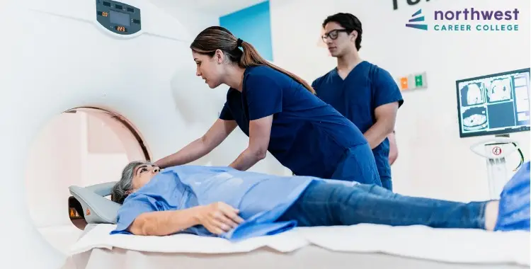
Practicing proper ways of protecting the patient during radiographic procedures is essential.
- Lead Aprons and Shields: A device made for shielding. (Note: many medical and dental facilities have begun discontinuing the use of these items over the past several years due to emerging research that suggests they no longer add protective value for patients as x-ray exposure techniques and equipment have improved)
- Positioning Aids: They enable one to get the correct alignment for patients, thus minimizing retake rates.
- Patient Education: Patients should be educated about the examination, probable risks, and the fact that they must be perfectly still at the time of exposure to prevent motion artifacts and increase the success rate with a single exposure.
Protect the Patients from Potential Hazards Associated with Radiation
Radiologic technologists are at risk of cumulative radiation doses because they work in imaging procedures regularly. Precautions include:
- Lead Shielding/Barriers: Barriers or lead shields embedded into walls create safe distances between the imaging room and the radiation source.
- Radiation Dosimeters: Always wear dosimeters to measure continuous radiation exposure and ensure that this does not accumulate into unacceptable doses.
Maintenance of Equipment and Quality Control
The radiographic equipment should be regularly maintained and quality controlled to ensure the best practices and safety.
- Routine Inspections: Regularly check and calibrate the X-ray machines to ensure proper exposure and function.
- Quality Assurance Programs: Quality assurance programs should include built-in tests and regular equipment performance checks.
- Repair at Once: Make prompt repairs of any faulty equipment or other flaws in the system that could cause unnecessary exposure or compromised image quality.
Training and Continuing Education
Radiologic technologists and other healthcare professionals must regularly undergo training and education to uphold radiation safety standards in their practice.
- Initial Training: Basic training in radiation safety, use of equipment, and protective measures.
- Continuing Education: Consists of regular training programs with information on the latest technologies, protocols, and best practices in radiation safety.
Conclusion
Radiation safety protocols for radiography are essential for patients and professionals to prevent potential risks associated with ionizing radiation. By following the principles of justification, optimization, and protective measures, radiographers can minimize their exposure and ensure radiographic technology’s safe and effective use.
Safety involves education, equipment maintenance, and strict adherence to safety standards in the radiographic environment. Prioritizing radiation safety allows for the utilization of radiography’s diagnostic power while safeguarding the health and well-being of everyone involved.


